Intro.
Blood vessels include arteries, veins, and arterio-venous connections (capillaries). All blood vessels have the same structure but differ in the relative thickness of each layer. This is the last lecture in the Cardio-vascular system.
Structure.
General structure.
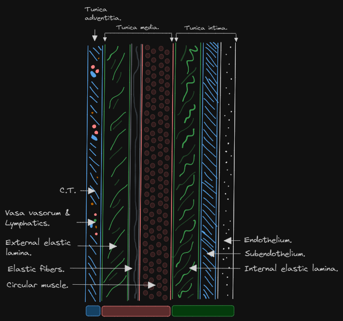
All blood vessels are composed of 3 tunics:
Tunica intima.
Innermost layer, consisting of 3 layers:
- Endothelium on BM.
- Subendothelium.
- Internal elastic lamina.
Tunica media.
Consisting of 3 layers:
- Circular smooth muscle.
- Elastic fibers.
- External elastic lamina.
Tunica adventitia.
Outermost layer, consisting of loose C.T which contains nerves, lymphatics, and vasa vasorum.
Arteries.
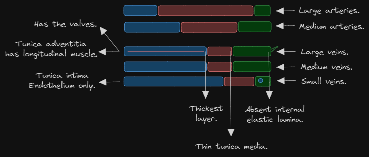
Can be classified into large arteries, medium arteries, and small arteries. They have thick walls and wide lumens.
Large arteries.
- Tunica intima 10% of total thickness.
- Tunica media 70% of total thickness (thickest layer).
- Tunica adventitia 20% of total thickness.
Medium arteries.
- Most of the arteries are medium-sized.
- Tunica intima has prominent elastic lamina.
- Tunica media mainly composed of circular smooth muscle.
- Tunica adventitia as thick as tunica media.
Small arteries.
(Isn’t explained, so skip.)
Specialized arteries. ⭐
- Cerebral arteries. (thin walled, prominent elastic tissue.)
- Coronary arteries. (thick walled, prominent elastic tissue.)
- Arteries of the lung. (thin walled, reduced muscle and elastic tissue.)
- Arteries of the penis. (develop longitudinally after puberty.)
- Umbilical arteries. (prominent muscular tissue, indistinct internal elastic lamina.)
- Roots of aorta and pulmonary arteries have some cardiac muscle fibers.
Veins.
Can be classified into large veins, medium veins, small veins. They have thin walls and wide lumen.
Large veins.
- Tunica intima is well developed, internal elastic layer is poorly defined (absent), and has valves.
- Tunica media is thin.
- Tunica adventitia is the thickest layer and has longitudinal smooth muscles.
Medium veins.
- Tunica intima has internal elastic layer which is poorly defined (absent).
- Tunica media is thin.
- Tunica adventitia is the thickest layer.
Small veins.
- Tunica intima endothelium only.
- Tunica media 1-2 layers of smooth muscle.
- Tunica adventitia is the thickest layer.
Connections between arteries and veins.
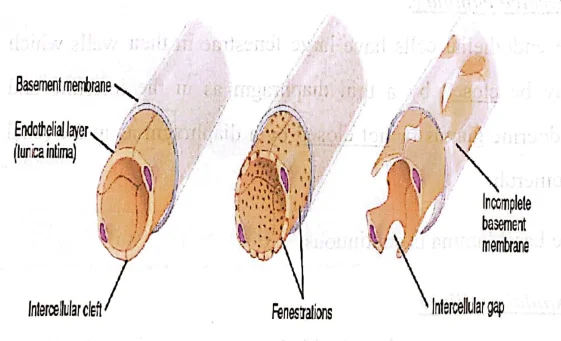
Capillaries.
Consist of:
- Single layer of endothelial cells. (represents Tunica intima.)
- Pericytes (represents Tunica media.)
- Thin layer of reticular fibers. (represents Tunica adventitia.)
They can come in many types such as:
- Continuous capillaries.
- No fenestrae.
- Strong basal lamina.
- Found in: muscles, C.T, and exocrine glands.
- Fenestrated capillaries.
- Have fenestrae which may be or may not be closed by diaphragm.
- Found in: intestines (diaphragmed), glomeruli (non-diaphragmed).
- Sinusoidal capillaries.
- Tortuous path and wide lumen.
- Fenestrated without diaphragms and has discontinuous basal lamina. (كل حاجه مخرمه)
- Found in: liver, spleen, and bone marrow.
Function of capillaries: ❌
- Permeability. (transfer of materials to and from tissue.)
- Metabolic function. (endothelial cells can metabolize a lot of substances.)
- Antithrombogenic. (blood does not stick to endothelial cells, preventing thrombi.)
Arterio-venous anastomosis. ❌
Consist of:
- They are arterioles that lose their internal elastic lamina and thicken their smooth muscle coat.
- Circular with narrow lumen.
- Regulates blood flow and temperature.