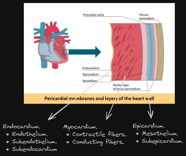
Layers of the heart.
Endocardium. (innermost layer.) ^b73221
Consists of 3 layers.
-
Endothelium.
- Single layer of simple squamous epithelium.
-
Subendothelium.
- Thin layer of loose connective tissue.
-
Subendocardium.
- C.T layer between endocardium & myocardium.
Myocardium. (middle muscle layer.)
- Thickest layer of the heart.
- Contains 2 types of muscle fibers:
- Contractile.
- Conducting.
Epicardium. (outermost layer.)
It is the Visceral Pericardium, consists of 2 layers.
-
Mesothelium.
- Layer of simple squamous epithelium sitting on thin C.T.
-
Subepicardium.
- C.T layer containing arteries, veins, and nerves.
Valves.
There are 4 valves in the heart:
- Tricuspid. → RA & RV.
- Pulmonary. → RV & Pulmonary Artery.
- Mitral. → LA & LV.
- Aortic. → LV & Aorta.
The valves are folds of the endocardium enclosing a central core of collagenous and elastic fibers. The inner edges of the valves at the side facing the ventricles are attached to the ventricles through papillary muscles through chordae tendinea.

Conducting systems of the heart.
The heart contains specialized fibers for transmitting impulses, these systems are:
-
SA node.
- The main pacemaker of the heart.
- Present under epicardium at junction of SVC and RA.
- Composed of modified non-contractile muscle cells and C.T.
-
AV node.
- Receives impulses from SA node.
- Present under endocardium at interatrial septum.
- Has same structure as SA node.
-
AV bundle.
- Originates from AV node.
- Formed of Purkinje fiber bundles separated by C.T.
- Branches at interventricular septum to right and left branches.
- Right branch: Moderator band.
- Left branch divides into 2 fascicles.
-
Purkinje fibers.
- Specialized cardiac cells that conduct impulses between the nodes and the surrounding ordinary muscle cells.
- These cells are larger, with abundant glycogen and a central deeply stained nucleus (or two.)
- They contain fewer myofibrils that are mostly central in position.
Cardiac muscle cells.
LM of cardiac muscle cells.
- Cardiac muscle is striated, involuntary muscle present in the heart’s myocardium.
- They are cylindrical, branching, and anastomose with other fibers.
- Cardiac muscle cells have a single central nucleus.
- The sarcoplasm of cardiac muscle cells is acidophilic and is interrupted between each cell with intercalated discs which are composed of facia adherens, desmosomes, and gap junctions.
(The sarcoplasm is the cytoplasm of muscle fibers.) (The sarcoplasmic reticulum is the smooth ER of muscle fibers.) (The sarcolemma is the cell membrane of muscle fibers.)
EM of cardiac muscle cells.
Cardiac muscle contains mitochondria and Golgi complexes among others such as:
T-tubules.
- They are invaginations of the sarcolemma into the sarcoplasm wider than those in skeletal muscle.
- They encircle the myofibrils at level of Z-line.
- T-tubule lumen is continuous with intercellular space, membrane is continuous with sarcolemma.
- Used to facilitate conduction of impulses along the fiber.
Sarcoplasmic reticulum.
- It is a special types of SER.
- Less developed and less regular than those in skeletal muscles, ends in small flattened portions that contact the T-tubules forming diads.
- Used to store Ca and releases Ca when wave of depolarization of sarcolemma arrives, causing contraction. Also actively transports Ca back into its cisternae after depolarization ends.
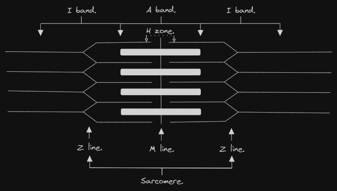
Myofibrils & myofilaments.
- Each myofibril is composed of myofilaments which are the contractile elements.
- Myofilaments exist in 2 types:
- Thin filaments. (actin)
- Thick filaments. (myosin)
- Arrangement of myofilaments gives cardiac muscle its striation by the alternating dark (A) and light (I) bands.
Sarcomeres are the structural contractile units of myofibrils, they are the portion of myofibril between 2 successive Z lines.
The dark band is called the A band and is formed of thick myosin filaments attached to M line. The light band is called the I band and is formed of thin actin filaments attached to Z line.
The H zone is a lighter area within the A band where myosin filaments are attached and is halved by the M line.
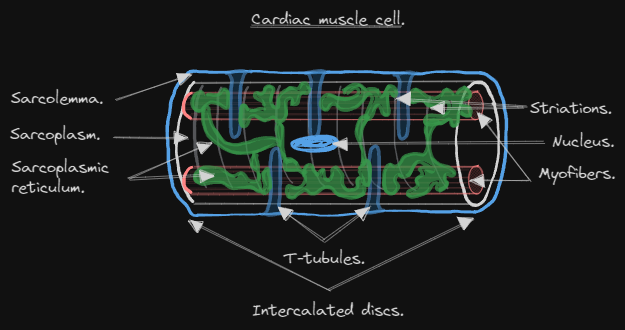
Intercalated discs.
They are the junctional complexes between 2 cardiac muscle cells. They use the following cellular junctions: Fascia adherens, Desmosomes, Gap junctions.
Fascia adherens.
- Forms the transverse portion of the intercalated disc. They hold the fibers together end-to-end.
Desmosomes.
- Forms both the transverse and the longitudinal portions of the intercalated disc. They bind the myocytes together.
Gap junctions.
- Mainly found in the longitudinal portion of the intercalated disc. They allow signals to pass from cell to cell allowing the heart to act as a syncytium.
Inclusions.
- Glycogen which is more abundant than in skeletal muscle cells indicating higher activity.
- Myoglobin which is rich in all cardiac muscle fibers giving them a red color.
- Lipofuscin which increases with age.
- Lipids.