Atria.
Right atrium.
Externally.
- Forms the sulcus terminalis.
- Has the right auricle.
Internally.
Divided into 2 parts separated by the crista terminalis:
- Sinus venarum. (smooth part)
- Posterior to crista terminalis.
- Receives blood from SVC and IVC.
- Has fossa ovalis with its margin, the limbus fossa ovalis.
- Atrium proper. (rough part)
- Anterior to crista terminalis.
- Derived from primitive atrium.
- Lined by pectinate muscles.
Orifices.
- SVC and IVC.
- Tricuspid valve.
- Venae cordis minimae.
Left atrium.
Externally.
- Forms posterior border.
- Has the left auricle.
Internally.
Divided into 2 parts:
- Inflow part. (smooth)
- Derived from pulmonary veins.
- Receives blood from pulmonary veins.
- Outflow part. (rough)
- Derived from embryonic atrium.
- Lined by pectinate muscles.
Orifices.
- Four pulmonary veins.
- Mitral valve.
Interatrial septum.
A solid muscular wall separating the right and left atria.
Ventricles.
Right ventricle.
Can be divided into:
Inflow part.
- It is at the inferior aspect of the ventricle.
- Covered by trabeculae carnae.
- Has papillary muscles connected to corda tendineae. (they prevent valve prolapse during systole.)
Outflow part. (Infundibulum.)
- It is at the superior aspect of the ventricle.
- Lacks trabeculae carnae.
- Derived from the bulbus cordis.
Left ventricle.
Can be divided into:
Inflow part.
- It is at the inferior aspect of the ventricle.
- Covered by trabeculae carnae.
- Has papillary muscles connected to corda tendineae. (they prevent valve prolapse during systole.)
(Basically the same as the inflow Part of the right ventricle.)
Outflow part. (Vestibule.)
- It is at the superior aspect of the ventricle.
- Lacks trabeculae carnae.
- Derived from the bulbus cordis.
(Basically the same as the outflow part of the right ventricle.)
Interventricular septum.
The Interventricular septum separates the two ventricles. It is composed of 2 parts: a superior membranous part, and an inferior muscular part which forms most of the septum. The muscular part has the same thickness as the left ventricular wall.
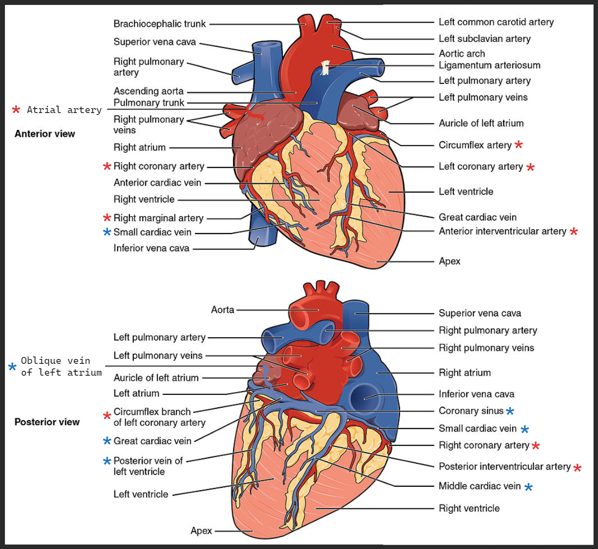
Vasculature of the heart.
”Vasculature of the heart” refers to the arterial supply and venous drainage of the heart.
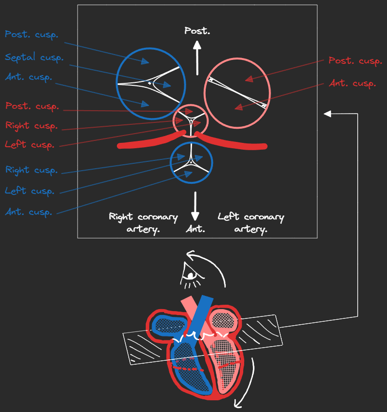
Arterial supply of the heart.
The arterial supply of the heart originates from the aortic sinuses that are found behind the anterior two cusps of the aortic valve (the right and left anterior cusps, giving rise to the right and left coronary arteries.) When the heart is relaxed, the back-flow of blood fills these sinuses, meaning the coronary arteries fill during cardiac diastole.
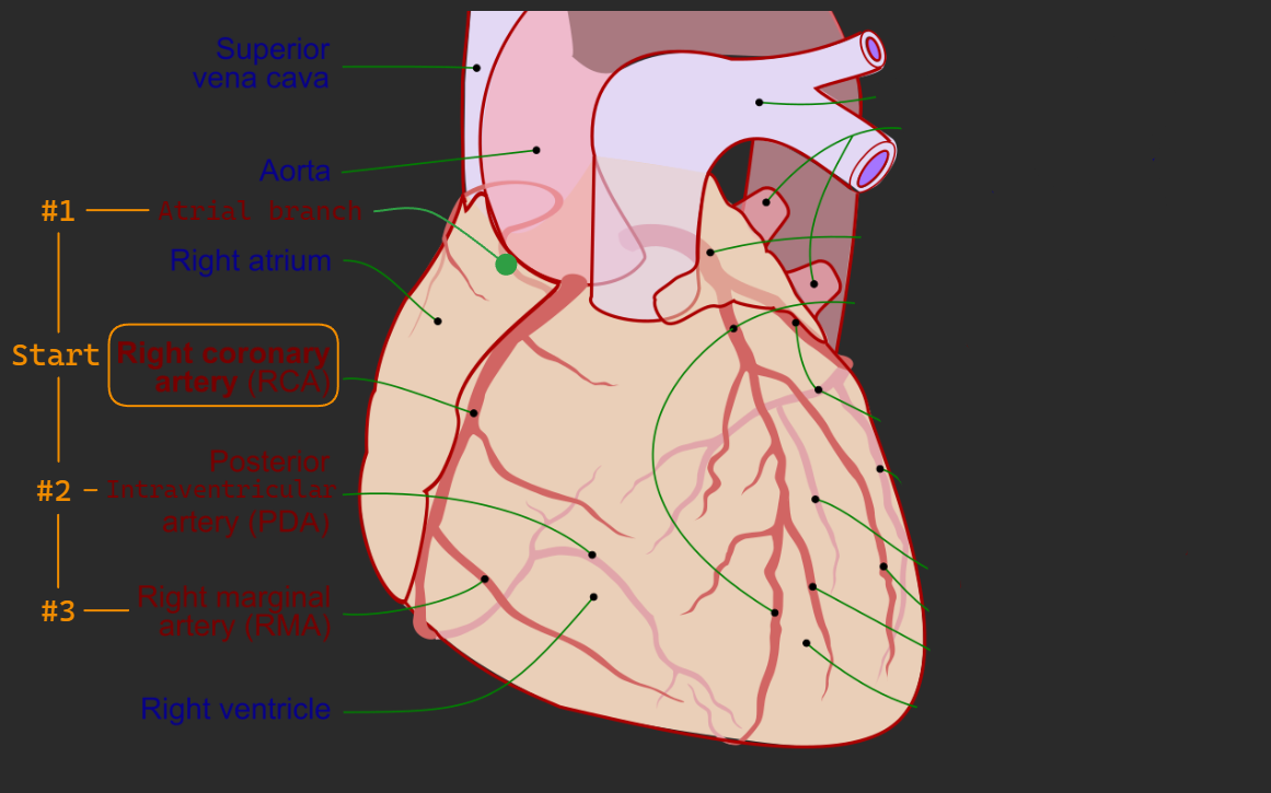
Right coronary artery.
- Origin: originates from the right aortic sinus in the ascending aorta.
- Pathway:
- Passes between the right atrium and pulmonary trunk.
- Enters coronary sulcus between right atrium and right ventricle.
- Moves posteriorly and continues in the sulcus on the diaphragmatic and posterior surfaces of the heart.
- Branches:
- Atrial branch: in the groove between right auricle and ascending aorta, loops around SVC and gives the SA nodal branch.
- Right marginal branch: at the inferior border of the heart, continues along inferior border towards the apex.
- Posterior intraventricular branch: in the posterior intraventricular sulcus on the diaphragmatic surface of the heart. Supplies a small branch to AV node.
- It supplies:
- Right atrium and ventricle. (Right coronary artery itself.)
- SA and AV nodes. (Right marginal artery → SA node.) (Posterior I.V. artery → AV node.)
- Interatrial septum and a portion of left atrium. (Posterior I.V. artery.)
- Posterior-inferior third of interventricular septum and a portion of posterior left ventricle. (Posterior I.V. artery.)
Left coronary artery.
- Origin: originates from the left aortic sinus in the ascending aorta.
- Pathway:
- Passes between left atrium and pulmonary trunk.
- Enters coronary sulcus.
- Divides to two branches behind the pulmonary trunk.
- Branches:
- Anterior interventricular artery: continues turning around to the left of pulmonary trunk and descends obliquely in anterior interventricular sulcus towards apex of heart. May supply one or two large diagonal branches (D1 and/or D2).
- The circumflex artery: continues posteriorly in the coronary sulcus to the diaphragmatic surface of the heart and ends before reaching the posterior interventricular sulcus. Branches into the left marginal arteries (M1 and/or M2).
- It supplies:
- Left atrium and ventricle. (Anterior I.V. artery and Circumflex artery.)
- AV bundle. (Circumflex artery.)
- Most of interventricular septum. (Circumflex artery.)
Venous drainage.
The coronary sinus receives venous drainage from 4 major sources and 2 minor sources:
Great cardiac vein.
- Origin: originates near the apex.
- Pathway: follows anterior interventricular sulcus into coronary sulcus where it turns left and joins the coronary sinus.
Middle cardiac vein.
- Origin: originates near the apex.
- Pathway: follows posterior interventricular sulcus towards coronary sinus.
Small cardiac vein.
- Origin: on the anterior surface of the heart.
- Pathway: passes around the right side of the heart to join coronary sinus.
Posterior cardiac vein.
- Origin: on the posterior surface of the left ventricle.
- Pathway: enters the coronary sinus directly or joins with the great cardiac vein.
Venae cordis minimae.
- Drains directly into right atrium and ventricle.
Oblique vein of left atrium.
- Ends in coronary sinus.