Intro.
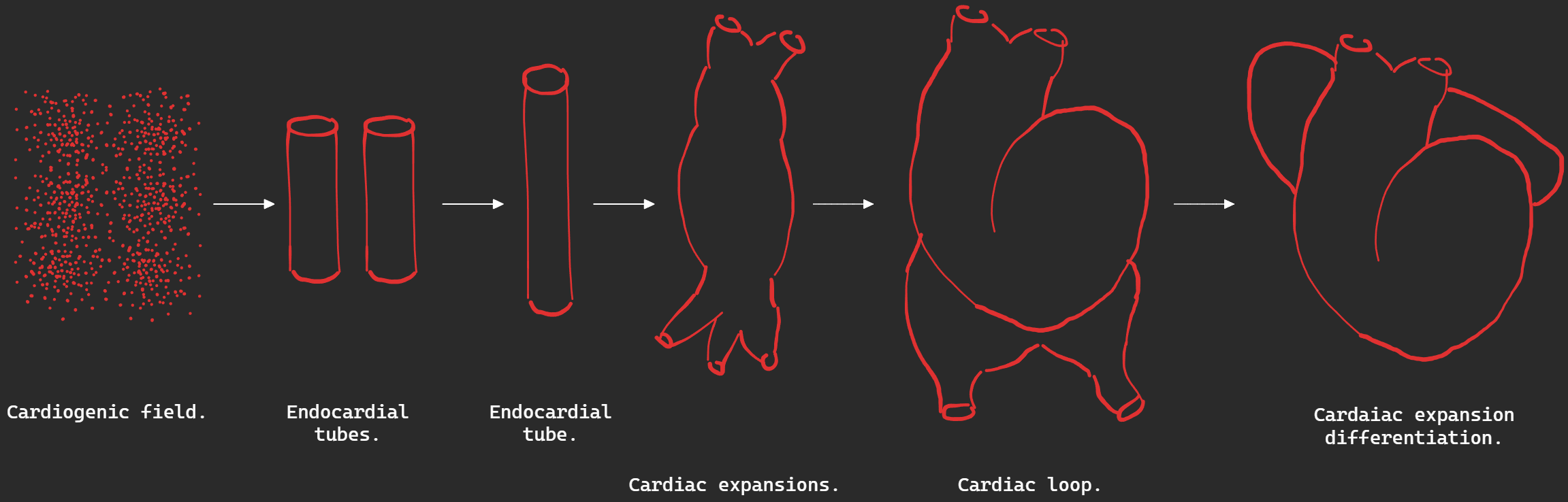
Development of the heart proceeds as follows:
- Cardiogenic field.
- Endocardial tubes. (tubes lined with endocardium)
- Primary heart tube.
- Cardiac expansions.
- Cardiac loop.
- Differentiation of loop.
Appearance cardiogenic field.
When the embryo requires more nutrition than is available by diffusion, it’s splanchnic mesodermal cells form angiogenic clusters around the cranial portion of the embryo.
Appearance of endocardial tubes and primary heart tube.
The clusters from the previous step form endothelial vessels which fuse to form right and left endocardial heart tubes cranially on either side of the embryo.
The embryo undergoes folding:
- Cranial folding brings the two tubes ventrally.
- Lateral folding brings the two endocardial tubes together to form one large endocardial tube.
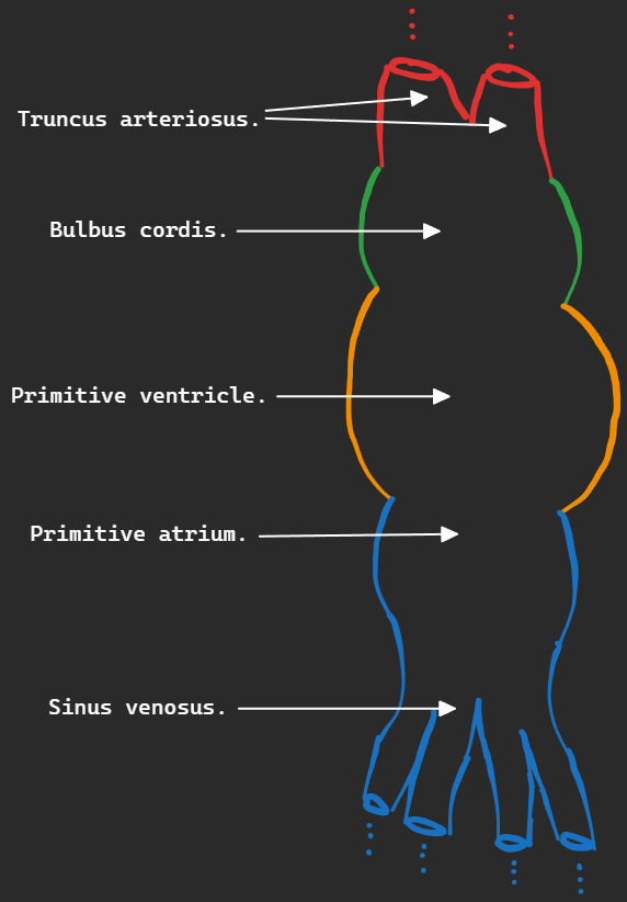
Cardiac expansions.
Cardiac tube already has constrictions indicating sectioning of the tube. From the bottom up (caudal→cranial) we have:
- Sinus venosus -⇒ Venous input of the heart and part of the atria.
- Primitive atrium -⇒ Adult atria.
- Primitive ventricle -⇒ Adult ventricles.
- Bulbus cordis -⇒ Most of the right ventricle.
- Truncus arteriosus -⇒ Pulmonary and aortic trunks.
Cardiac loop and its differentiation.
As the cardiac tube elongates, it loops around itself first in a “C” shape, then in an “S” shape due to its fixed ends, disproportional growth between it and the pericardial sac and between different parts of the tube.
Development of the sinus venosus.
Sinus venosus collects blood from 3 veins on both sides:
- Left and right Umbilical veins.
- Left and right Vitelline veins.
- Left and right Common cardinal veins.
At first, left and right sides are equal. Blood flow shifts from left to right, as well as stoppage of umbilical flow causing obliteration of left and right umbilical veins and the left vitelline vein. Left side now only has the common cardinal vein which becomes the oblique atrial vein and the entire left side becomes the coronary sinus.
The right side enlarges due to increased blood flow and is absorbed into the right atrium to form it’s posterior smooth part.
- Left common cardinal vein -⇒ coronary sinus and oblique atrial vein.
- Right common cardinal vein -⇒ lower part of SVC.
- Right vitelline duct -⇒ opening of IVC.
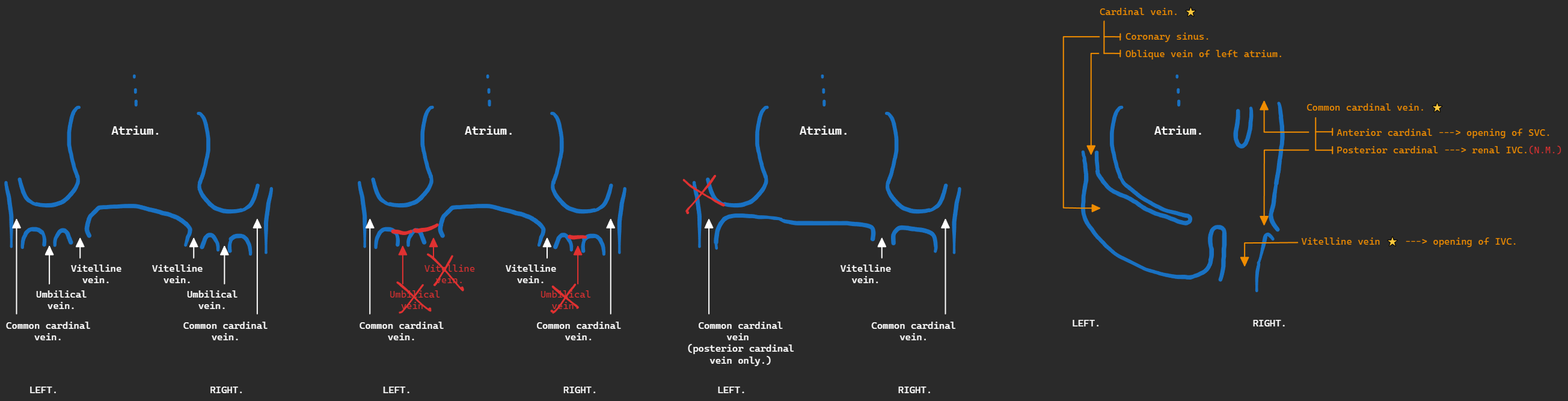
The sinoatrial orifice (the opening between the sinus venosus and the atrium) is guarded by 2 valves:
- Left venous valve and septum spurium fuse with interatrial septum.
- Upper right venous valve forms cristae terminalis.
- Lower right venous valve forms valves of IVC and coronary sinus.
Now the heart looks like this after differentiation of the heart loop:
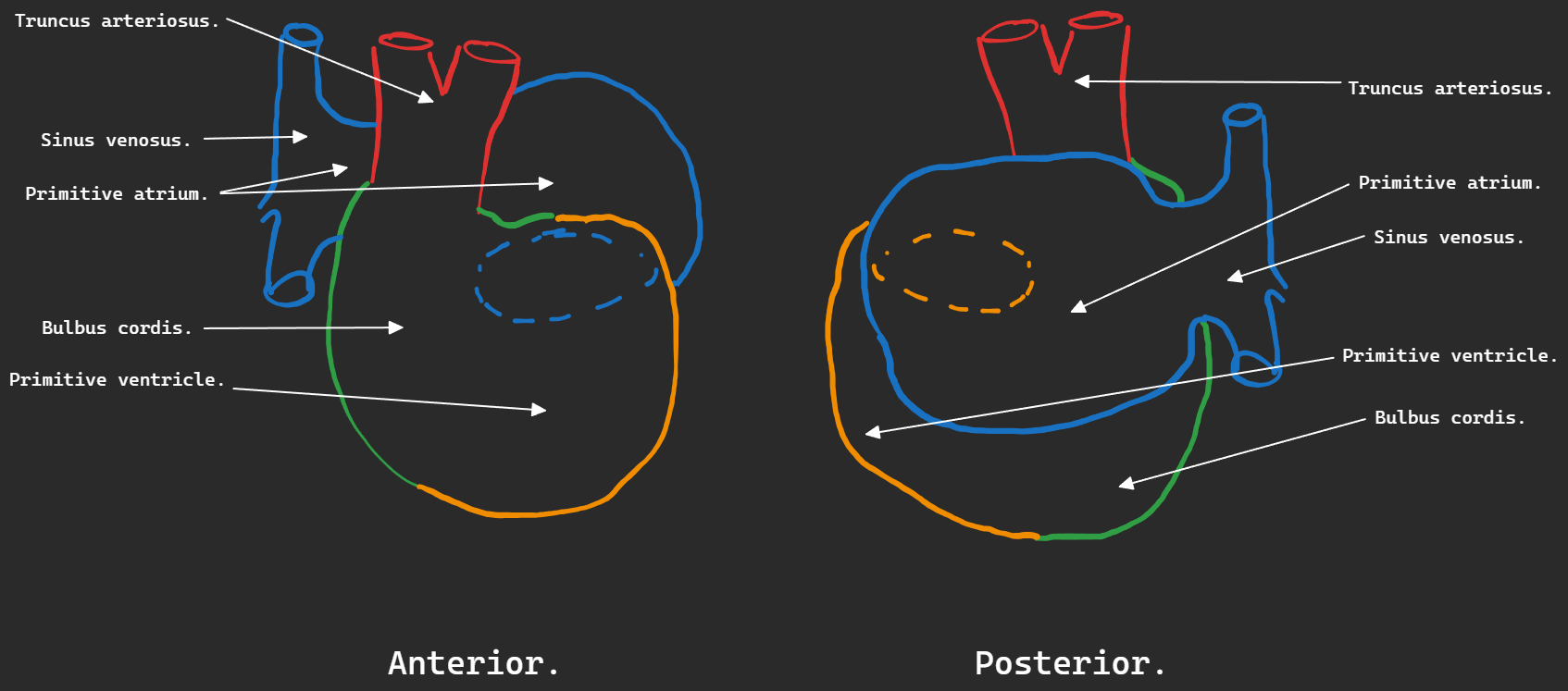 (Note: the connection between the common atrium and common ventricle gradually moves upwards.)