Intro.
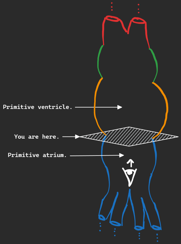
Last lecture we discussed the development of the sinus venosus. The next step in heart creation is to create septa to separate the atria from the ventricles. From these septa we will also create valves. First lets look at the interatrial septum and the atria, then after that we’ll look at the ventricles and the interventricular septum.
Interatrial septum and atrial development.
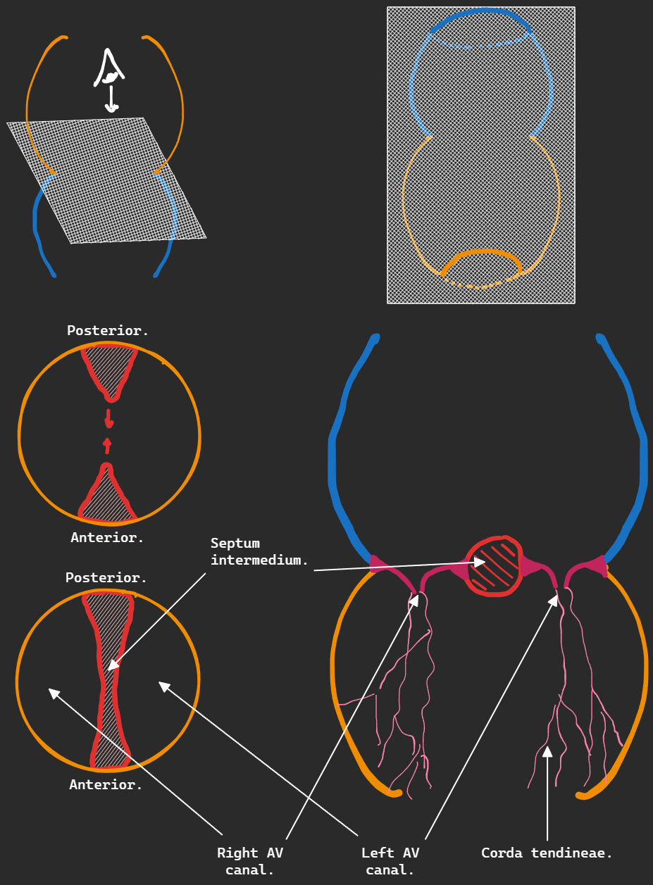
Septum intermedium.
The development of the interatrial septum begins with the separation of the atrioventricular canal into left and right AV canals by the anterior and posterior endocardial cushions. These cushions fuse together forming the septum intermedium. Off of which the tricuspid and mitral valves and their corda tendineae attach.
Septum primum.
After the appearance of the septum intermedium and subsequent folding of the heart tube, the septum primum appears from the top of the common atrium and descends down, creating the foramen primum, eventually reaching the septum intermedium (foramen primum closure.) The cranial (upper) part of the septum primum break causing the formation of the foramen secundum.
Septum secundum.
After the appearance of the foramen secundum, the septum secundum starts forming at the top of the common atrium, on the right side of the septum primum. The edge of the septum secundum overlaps with the foramen secundum, leaving only a tiny slit for the blood to pass through, bypassing the pulmonary circulation. The slit is called the foramen ovale.
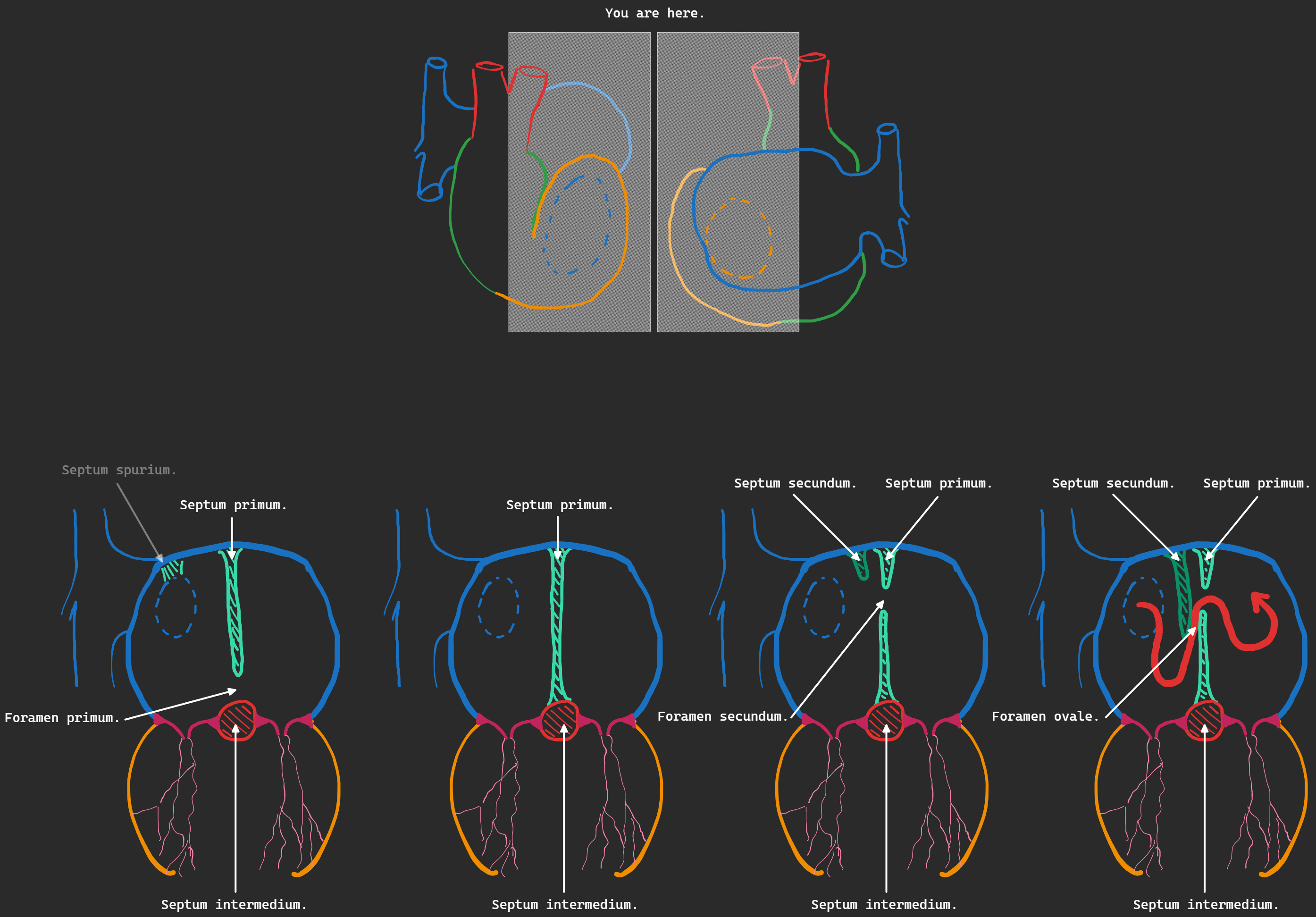
Formation of the interatrial septum.
Right atrial blood pressure decreases due to a decrease in placental circulation. Left atrial blood pressure increases due to increased pulmonary venous return. These 2 factors cause the foramen ovale to close, forming the fossa ovale. And causing the septum primum to be pushed towards the septum secundum forming the interatrial septum. ⭐ The septum secundum also forms the limbus fossa ovale (the raised edge of the fossa ovale.)
Components of the atria.
Right atrium:
- Right half of primitive atrium.
- Absorbed right horn of sinus venosus.
- Upper part of right atrioventricular canal.
Left atrium.
- Left half of primitive atrium.
- Absorbed pulmonary veins.
- At first a common PV is created by fusion of all 4 PVs, the common PV is then absorbed into the left atrium.
- Upper part of left atrioventricular canal.
Defects of interatrial septum.
Persistent foramen ovale.
Due to incomplete fusion of the septum primum and septum secundum.
Premature closure of foramen ovale.
Leads to hypertrophy of right side of the heart and an underdeveloped left side.
Foramen secundum defect.
Due to defective development of septum secundum or resorption of septum primum.
Common atrium.
Due to complete failure of septum primum and septum secundum development.
Interventricular septum and ventricular development.
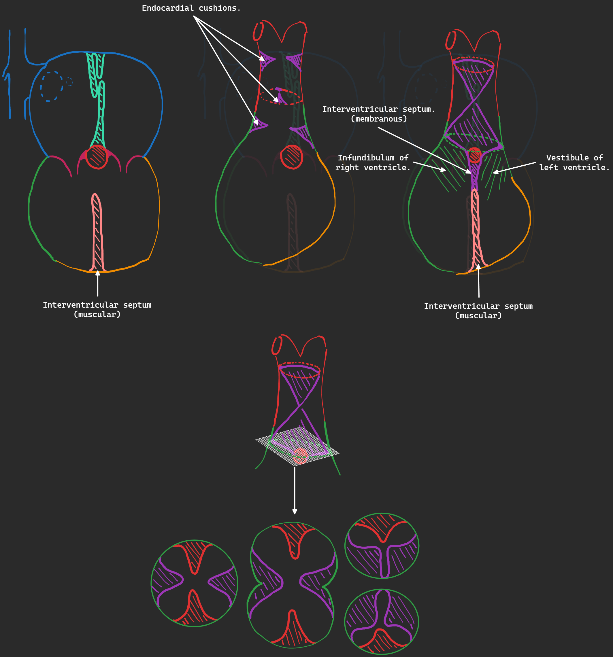
First, similar to what we did in interatrial septum, the interventricular septum begins from the floor of the common ventricle and extends upwards, creating the interventricular foramen.
Ridges appear longitudinally in the bulbus cordis and truncus arteriosus, they extend towards each other until bulbus cordis and truncus arteriosus are split in two (the bulbus cordis giving rise to the infundibulum of right ventricle and vestibule of left ventricle.)(the truncus arteriosus giving rise to the pulmonary trunk and aorta.)
The ridges also extend towards the muscular interventricular septum, creating the membranous interventricular septum (closing the interventricular foramen.)
Components of ventricles.
Right ventricle.
- Right part of primitive ventricle.
- Right part of bulbus cordis (infundibulum.)
Left ventricle.
- Left part of primitive ventricle.
- Left part of bulbus cordis (vestibule.)
Defects in interventricular septum.
Membranous interventricular septum defect (VSD.)
Due to failure of formation of membranous IVS.
Muscular interventricular septum defect (VSD.)
Due to failure of formation of muscular IVS.
Common ventricle.
Due to failure of membranous and muscular IVS.